Attention: Restrictions on use of AUA, AUAER, and UCF content in third party applications, including artificial intelligence technologies, such as large language models and generative AI.
You are prohibited from using or uploading content you accessed through this website into external applications, bots, software, or websites, including those using artificial intelligence technologies and infrastructure, including deep learning, machine learning and large language models and generative AI.
Nephroblastoma (Wilms' Tumor)
- Constitute ~85% of childhood renal cancers.
- Age: 2-4 years; extremely uncommon in children <6 months and >6 years.
- Associations: WAGR syndrome, Beckwith-Wiedemann syndrome, and Denys-Drash syndrome.
- Mostly presents as abdominal mass. (Detected by parents).
- Bilateral in 5% of cases.
- Radiology: calcification is uncommon (vs. neuroblastoma, which is often calcified).
- Gross:
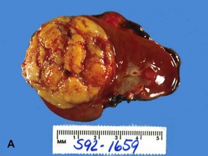
- Usually large with solid soft gray tan cut surface and seemingly well-circumscribed borders (image A).
- Hemorrhage and necrosis are common.
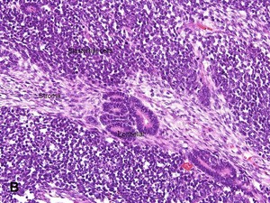
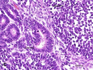
- Microscopic: 3 distinct elements:
- Blastema: sheets of densely packed primitive small cells with scant cytoplasm and darkly staining nuclei ("small round blue cell tumor") (image B) & (image C).
- Epithelial: small tubules or cysts lined by primitive or mature columnar or cuboidal cells.
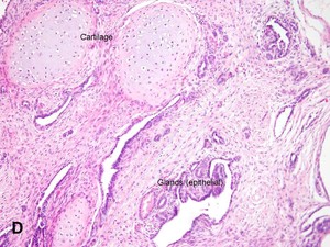
- Stroma: spindle cells or may differentiate along the lines of any soft tissue such as fibroblasts, smooth muscles or skeletal muscles, cartilage (image D); loose myxoid and fibroblastic spindle cell stromas are the most common.
- Most tumors though have biphasic or monophasic growth.
- Immunohistochemistry: WT1+ in blastemal and epithelial components.
- Overall survival is good (>90%).
- Unfavorable prognostic factors are high stage and presence of diffuse anaplasia.
- DDX:
- Other small round blue cell tumors including neuroblastoma (WT1-).
advertisement
advertisement


